To develop an automated vertical cup-to-disc ratio (vCDR) estimation system that can work across different types of retinal fundus images, including standard fundus images, ultra-widefield images and smartphone-based images.
The cup-disc ratio (CDR) is used to assess the optic nerve head and evaluate the risk or presence of glaucoma, a group of eye conditions that can lead to blindness due to damage to the optic nerve. The "cup" refers to the central depression in the optic nerve head, and the "disc" is the entire optic nerve head. In glaucoma, increased intraocular pressure can cause the optic nerve fibers to be damaged, leading to an enlarged cup and, consequently, a higher CDR. Regular monitoring of the CDR is essential for early detection and management of glaucoma to prevent vision loss.
For more information on this study, you can visit this article on the TVST journ
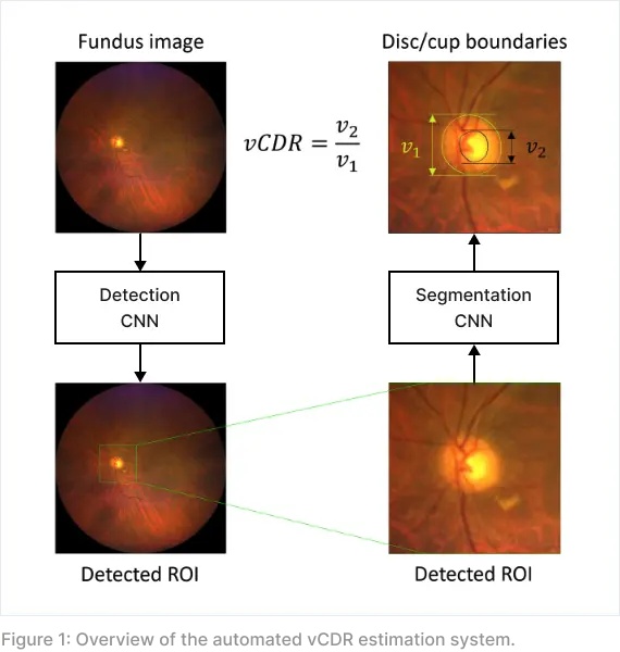
Method overview
The system is powered by deep neural network, where two sequential convolutional neural networks (CNNs) were trained to identify the region of interest around the optic disc and delineate the boundaries of optic disc and optic cup for vCDR calculation. An overview of the automated system is illustrated in Figure 1.
Validation on smartphone-based images
To assess the performance of the system, a total of 300 smartphone-based fundus images were collected from 151 patients at the AMK specialist center using the oDocs Nun IR camera. A demonstration of the image acquisition process is given in Figure 2.

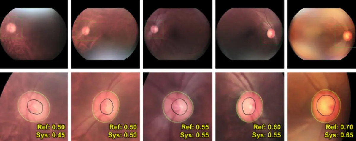
The vCDRs computed by the automated system were compared against the reference values annotated by a team of optometrists at TTSH. The automated system performs reasonably well in smartphone images of varying quality. Some sample outputs are given in Figure 3.